Immuno-positron emission tomography (PET) imaging can provide early insight into a tumor’s response to targeted therapy, allowing physicians to select the most effective treatment for patients who have cancer. The new research was published in the March issue of The Journal of Nuclear Medicine.
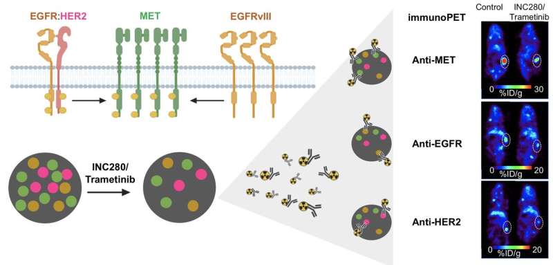
The research showed that immuno-PET successfully visualizes changes in different cancer receptors (receptor tyrosine kinases, or RTKs) within tumors during targeted therapies. This gives physicians a tool that can be used to evaluate the effectiveness of a treatment soon after its administration.
“When healthy cells turn into cancer cells, there is a disruption in the RTK signaling. This makes RTKs a valuable therapeutic and imaging target,” said Patricia Pereira, Ph.D., a research associate at Memorial Sloan Kettering Cancer Center in New York, New York. “Techniques that allow for real-time monitoring of RTK dynamics, such as immuno-PET, could be very beneficial in informing treatment choice and predicting response.”
Immuno-PET uses a “tracer” to follow an antibody directed to a specific tumor. This allows physicians to obtain images of events happening at the tumor site and provides information into whether the tumor responds to the treatment. The physician can then visualize how the tumor is responding.
In this study, researchers used immuno-PET and three different antibodies to visualize three RTKs (MET, EGFR, and HER2) in a kidney tumor. Their results confirmed that immuno-PET visualizes RTKs in ways that determine the level of protein within a tumor. After administering a treatment, immuno-PET can detect changes in RTK levels that indicate whether a tumor is responsive to that treatment.
Source: Read Full Article


