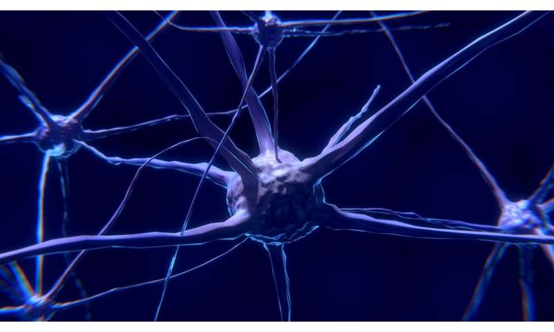
A study team led by MedUni Vienna’s Center for Brain Research has charted how cell types that form neuroendocrine command centers that control virtually all aspects of bodily metabolism evolve during brain development. The study, which employed the most advanced tools to distinguish cells at the molecular level, showed entirely unexpected origins and developmental programs for many nerve cells, and describes how millions of neurons assemble into a precisely knit network by birth. They have also found key molecular signatures, called transcription factor networks, whose impairment was directly linked to obesity, post-traumatic stress and sleep deficits (narcolepsy/insomnia). Understanding the developmental design of the brain area that hosts highest-order command cells for key hormonal systems will advance the understanding of virtually all neuroendocrine diseases with developmental origins.
The study, published in the journal Nature, examines how the hypothalamus develops in mouse models and human disease-related genome-wide association data. The hypothalamus contains a kaleidoscope of neuroendocrine command neurons that orchestrate hormonal responses against virtually any peripheral stimulus one experiences throughout life. Hypothalamic centers are central to regulating, among other things, stress, body temperature, sleep and day-night cycles, reproduction, food and fluid intake and body weight. A key difficulty the researchers faced was that the hypothalamus evolved to house its many cell types in a minimal brain volume, thus undoubtedly being the most heterogeneous and functionally complex area of the brain.
The study primarily relied on single-cell RNA sequencing, the most advanced tool kit to molecularly profile cell types. Data from tens of thousands of cells were integrated to understand when and where nerve cells are born, how they find their final positions and mature into neuronal types that can orchestrate hormonal responses, and by which mechanisms they sense bodily signals and communicate those to higher brain centers. As a result, they were able to not only rationalize the organization of all neuronal types that have been shown to exist in the adult hypothalamus over the past century but also discovered hitherto unknown neurons.
The research team have particularly looked at dopamine neurons, which inhibit the release of prolactin, the hormone regulating fertility and milk production after pregnancy once released from the pituitary. They have found that these cells arise from precursor cells that rather switch their identities than being dedicated to generate a single type of neuron. Subsequently, they become one of at least nine subtypes of dopamine cells. It was also shown that the program of differentiation for dopamine cells is regulated by a unique set of molecules, transcription factors, whose mutations in humans induce obesity and abnormal stress responses. Thereby the concept that neurons carrying the same neurotransmitter (that is, the signal molecule used to communicate with other nerve cells) are functionally similar or even identical was broken and replaced by the concept of “molecular heterogeneity coding for functional diversity” through the coincident use of neurotransmitters, hormones and other signaling molecules.
Unexpected complexity
“Demonstrating the many cell types in the hypothalamus is a breakthrough, because it will provide a cellular template to study hormonal functions and neuroendocrine and metabolic diseases,” explains principal investigator Tibor Harkany, head of the Department of Molecular Neurobiology at MedUni Vienna’s Center for Brain Research, who also holds a professorship at Sweden’s Karolinska Institutet. “We expect nothing less than the discovery of novel modes of brain-periphery communication, perhaps even by as yet unknown hormones, in the not-too-distant future.”
Clearly, the many cell types defined in this study will require experimental follow-up to distinguish their specific functions, and to identify both communication partners in other brain areas and sensitivity to peripheral hormones. “This study is a milestone, because it rationalized one of the most complex brain areas that controls more outputs fundamental to sustain life than any other we know of,” says Tomas Hökfelt of the Karolinska Institutet and adjunct professor at MedUni Vienna. “Thereby, we will be able to place many of the exquisite early findings that focused on individual cell types into systems-wide contexts and estimate their relative contributions to various neuroendocrine diseases.”
Hope for regenerative medicine
Source: Read Full Article


