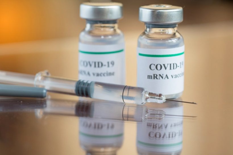
In a recent study published in Nature Biotechnology, researchers reported on the automatic printing of microneedle patch (MNP) severe acute respiratory syndrome coronavirus 2 (SARS-CoV-2) messenger ribonucleic acid (mRNA) vaccines.

Image Credit: chatuphot/Shutterstock.com
Background
Unvaccinated individuals in developing nations are at an elevated risk of recurrent SARS-CoV-2 infections, which have caused unprecedented morbidity and mortality globally. Mass-scale coronavirus disease 2019 (COVID-19) vaccination in community settings is hindered by insufficient cold-chain logistics and a lack of trained healthcare personnel. Decentralized, local-level manufacture of thermostable mRNA vaccines using an MNP format could provide a probable solution.
Intradermally (ID)-delivered MNPs are self-administrable, cause less pain than intramuscular (IM) delivery, do not produce sharps waste, and are stable for several months. Pfizer-BioNTech’s and Moderna and COVID-19 mRNA vaccines, comprising lipid nanoparticles (LNPs), have effectively prevented COVID-19 severity outcomes.
About the study
In the present study, researchers used microneedle vaccine printing (MVP) to fabricate thermostable COVID-19 mRNA vaccines in an LNP vehicle.
To fabricate MNP by microneedle vaccine printing, modular inks comprising mRNA-loaded LNPs and a dissolvable stabilizing polymer blend were used. The ink was optimized for high bioactivity by screening formulations in vitro and required to be consistently dispensable. Following automatic dispensing, a vacuum was applied through the polydimethylsiloxane (PDMS) mold for loading the microneedle with the viscous vaccine-polymer solution.
Subsequently, the molds were placed in drying stations for rapid drying. The team measured the loading times for several parameters associated with MVP vaccine fabrication and design. LNPs of 147.0 nm diameter, which encapsulated mRNA encoding for firefly luciferase (fLuc), were mixed with various water-soluble polymers and dried.
Thermogravimetric analysis was performed to evaluate drying time, and drying rates for various drying strategies were compared by linear regression modeling. The drop area and MNP coverage of the polymer solution were also evaluated. MNPs on an ultrathin acrylic backing, produced using the standalone device, were imaged using scanning electron microscopy (SEM). The throughput of the microneedle patch was assessed in terms of tray lengths and the duration required for drying.
The shelf stability of the resulting MNPs was assessed using a model mRNA construct. The single-step and double-step vaccine loading efficiencies were assessed. The efficiency of protein and deoxyribonucleic acid (DNA) loading in manually produced MNPs was evaluated. Protein expression was measured in Henrietta Lacks (HeLa) cells following transfection with re-dissolved polymers with similar protein expression as suspended LNPs. Luminescence was observed after six hours of MNP application with cKK-E12 or lipid 5.0-comprising LNPs in C57BL/6 mice.
The team compared the administration of cKK-E12-comprising LNPs encoding fLuc messenger ribonucleic acid via the IM route, printed MNP, and manually fabricated MNP. Further, mice were vaccinated with encapsulated mRNA coding for SARS-CoV-2 spike (S) glycoprotein receptor-binding domain (RBD) via MNP, followed by an MNP boost 4.0 weeks later. Serum anti-S RBD immunoglobulin G (IgG) titers were measured using electrochemiluminescence anti-S RBD binding assays.
Results
The MNPs showed a ≥6.0 month-shelf stability at room temperatures. Vaccine loading efficiency and MNP dissolution indicated that efficacious, microgram-level dosages of LNP-encapsulated mRNA could be administered by an MNP. Vaccinations in mice with SARS-CoV-2 spike RBD stimulated durable immunological responses comparable to those induced by IM administration. The SEM analysis showed images of MNPs with conical and pyramid geometries. The efficiencies of protein and DNA and protein loading in manually fabricated and MVP-fabricated MNPs were comparable.
The heat maps showed consistent DNA loading in microneedle patches across 10.0 × 10.0 mold trays, where each square represented a distinct patch. Microneedles dissolved and delivered mRNA-LNP vaccines to the intradermal space. Pyramid MNPs comprising a 1:1 blend of polyvinylpyrrolidone (PVP) and polyvinyl alcohol (PVA) were stronger and stiffer than conical MNPs. Moreover, 36.0% and 8.0% of the pyramidal and conical MNP volume dissolved within 10 minutes, respectively, indicating that pyramidal MNPs enabled greater vaccine delivery.
PVP:PVA stabilized mRNA-LNPs in MNPs for high protein expression six months post-storage at room temperatures. MVP-produced MNPs had adequate mechanical properties and successfully penetrated the porcine epidermis ex vivo. Testing manually fabricated MNPs In vivo showed immune responses comparable to that induced by IM vaccination. After 3.0 weeks of boosting, the geometric mean titers (GMTs) were similar between IM and MNP for the SARS-CoV-2 RBD mRNA vaccine. Despite similar anti-S RBD IgG titers for IM and MNP with SARS-CoV-2 RBD mRNA, some differences were noted between IM administration of mRNA-LNP suspension and ID delivery of mRNA-LNPs using MNPs.
Lipid 5.0-incorporating mRNA-LNPs showed greater protein expression than cKK-E12, indicative of MNP sensitivity to ionizable lipids. Modeling data indicated that the mRNA-LNP SARS-CoV-2 vaccine could be formulated into a 2.0-cm-square MNP for human application. The model predicted that 108 and 360 of the pyramid MNPs could deliver the complete dose of Pfizer-BioNTech and Moderna vaccines, respectively. Loading of increasingly viscous inks required <20.0 minutes, and LNP loading efficiency was enhanced by using small-sized MNPs, reducing the Marangoni effect and maximizing circularity. The first-generation printer manufactured 100.0 patches in 2.0 days, the efficiency of which could be enhanced by vertical stacking of mold trays or continuous fabrication of MNPs by de-gassing the trays prior to dispensing.
Conclusion
Overall, the study findings highlighted the development of an MVP to fabricate dissolvable MNPs loaded with LNP-encapsulated mRNA vaccines or other cargo with microliter-scale precision through automated fabrication processes, minimizing user interactions. The vaccines could be stored at room temperature and used months after fabrication, facilitating use in resource-limited settings and allowing cost-effective stockpiling of vaccines to improve preparedness against future outbreaks.
- vander Straeten, A., Sarmadi, M., Daristotle, J.L. et al. A microneedle vaccine printer for thermostable COVID-19 mRNA vaccines. Nat Biotechnol (2023). doi: https://doi.org/10.1038/s41587-023-01774-z https://www.nature.com/articles/s41587-023-01774-z
Posted in: Medical Science News | Medical Research News | Disease/Infection News
Tags: Alcohol, Biotechnology, Cold, Coronavirus, Coronavirus Disease COVID-19, covid-19, DNA, Electron, Electron Microscopy, Epidermis, Ex Vivo, Glycoprotein, Healthcare, heat, Immunoglobulin, in vitro, in vivo, Lipids, Luciferase, Microscopy, mold, Mortality, Nanoparticles, Pain, Polymers, Protein, Protein Expression, Receptor, Respiratory, Ribonucleic Acid, SARS, SARS-CoV-2, Severe Acute Respiratory, Severe Acute Respiratory Syndrome, Syndrome, Transfection, Vaccine

Written by
Pooja Toshniwal Paharia
Dr. based clinical-radiological diagnosis and management of oral lesions and conditions and associated maxillofacial disorders.
Source: Read Full Article
