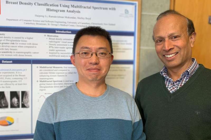
A University of Canterbury (UC) student has come up with a new computerized method of reading mammograms that could help radiologists detect warning signs of breast cancer.
Haipeng Li is about to complete a Ph.D. in Software Engineering at UC after spending the last three years working on computational algorithms that can automatically read and analyze mammogram X-rays.
The algorithms he has developed, with UC Professor Ramakrishnan Mukundan and radiologist Dr. Shelley Boyd at Pacific Radiology in Christchurch as his supervisors, have been shown to accurately detect two markers linked to increased risk of breast cancer.
He hopes the research will eventually help radiologists identify cancers at an early stage when they can be treated more successfully.
“Early detection through routine mammograms plays an important role in preventing breast cancer deaths,” Li says. “But reading and interpreting suspicious regions in mammograms is repetitive and challenging work.
“The algorithms I’ve been working on are designed to make it easier for radiologists to pick up two biomarkers for breast cancer—microcalcifications and mammogram density. Tiny calcium deposits in the breast and dense breast tissue are both indicators of a higher risk of developing breast cancer.”
Breast cancer is one of the most common cancers in women with over 1.5 million diagnosed worldwide each year.
In New Zealand, there are more than 3000 diagnosed cases and more than 600 deaths every year.
Mammography is a widely accepted imaging method used for routine breast cancer screening around the world and it can detect about 80%–90% of breast cancer cases before symptoms appear.
Li’s new techniques using advanced machine learning algorithms have shown higher accuracy rates in automatically detecting microcalcifications and dense breast tissue compared to other computerized tools being used by radiologists internationally.
His model, using texture features extracted from mammogram images, provides more specific position information on the location of microcalcifications.
“I also tested over 700 mammogram images for mammogram density and obtained classification accuracy of 92% and 87% on two public mammogram datasets,” Li says. “Very few computerized classification systems being used overseas have accuracy rates of more than 80% so it’s a very significant achievement.”
He says his aim is to provide a “secondary perspective” to support radiologists’ work and make it easier for them to interpret images, rather than computers replacing people.
“Knowing that the work I’m doing will contribute to people’s health outcomes makes it more meaningful for me. It’s using artificial intelligence to help human beings and change our lives for the better.”
Li, who worked as a university lecturer in China for a decade before coming to New Zealand in 2018, hopes his proposed automated algorithms will feed into computer aided detection and diagnosis (CAD) systems in the future.
He says further improvements and evaluations are needed before the tool could be used in a clinical setting.
“We’re aiming to reach 98% accuracy. Some improvements are needed in the current algorithms to extract more detailed image information for specific biomarkers and I hope to work more closely with local hospitals and radiologists to achieve even better results.”
UC Computer Science and Software Engineering Professor Mukundan, who is the principal supervisor for Li’s research, says computerized approaches are increasingly being used internationally for interpretation and analysis of diagnostically relevant features in X-rays.
“Li’s research makes significant contributions to this field by performing a comprehensive texture anaysis of mammographic features for microcalcification detection and breast density estimation.”
Radiologist Dr. Boyd says detection of microcalcification from mammograms is a major part of breast cancer detection. Early treatment can prevent progression to invasive and more aggressive forms of the disease. It is also useful to have local research relating to breast density, which is a “hotly debated” topic in the breast imaging world at present, she says.
Source: Read Full Article
