Scientists at the University of Virginia School of Medicine have identified a potential way to head off heart attacks and strokes by strengthening the fibrous caps overlying atherosclerotic plaques that naturally accumulate inside our arteries. These fatty lesions can rupture, triggering blood clots that cause disability or death.
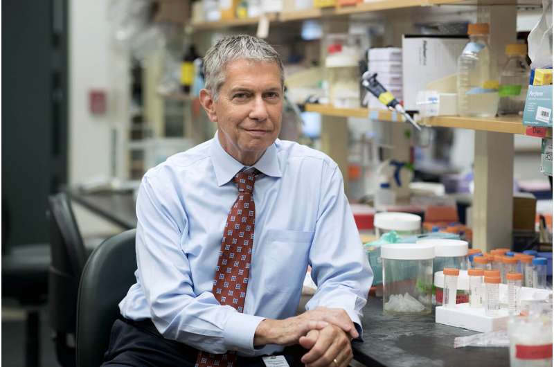
New research from the lab of UVA researcher Gary K. Owens reveals surprising new information about the makeup of the protective caps our bodies build over these lesions, and about the factors that determine their stability. The study supports recent findings that certain types of inflammation might actually help stabilize plaques. Doctors may one day be able to use these insights to strengthen the caps and prevent the plaques from rupturing.
“These studies redefine our understanding of both how the caps form and what makes them strong,” said Owens, the head of UVA’s Robert M. Berne Cardiovascular Research Center and a member of UVA’s departments of Molecular Physiology and Biological Physics and of Internal Medicine (Division of Cardiology). “These studies were completed by a large team of highly talented investigators from UVA and abroad but led by three outstanding trainees from my laboratory, including co-first authors Alexandra Newman, Vlad Serbulea and Dr. Richard Baylis.”
Understanding Atherosclerotic Plaques
Unstable atherosclerotic plaques account for the majority of heart attacks and a large fraction of strokes, making these lesions the leading cause of death worldwide. The protective caps our bodies create over these lesions act like a patch on a tire, preventing them from rupturing and triggering catastrophic blood clot formation, which, in blood vessels supplying the heart or brain, can cause a heart attack or stroke. Therefore, improving our understanding of how the cap forms is of major clinical importance. “Despite decades of research, little is known regarding the factors and mechanisms that promote formation and maintenance of a stable fibrous cap,” the UVA researchers write in a new scientific paper outlining their findings.
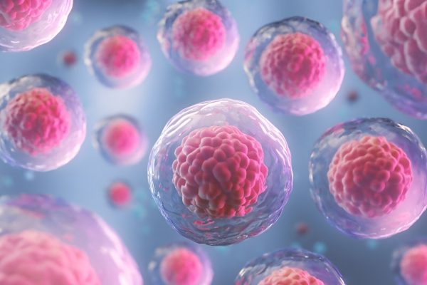
This work from Owens and his team helps change that, offering unexpected insights into the caps’ composition and origins. Scientists have thought that the caps were derived almost exclusively from smooth muscle cells, but Owens’ findings reveal that there is a “tapestry” of different cell types involved. “For years we assumed that most of the protective fibrous cap cells were of smooth muscle cell origin because that’s what they look like under the microscope,” Newman said, adding, “Advanced techniques show us how dynamic this structure really is.”
Baylis noted that “having multiple cell types contribute to the fibrous cap likely make this critically important structure more robust and resistant to plaque rupture.”
Up to 40% of the fibrous cells in the cap in lab mice come from sources other than smooth muscle cells, the researchers found. In advanced human lesions, approximately 20% to 25% of the fibrous cap cells came from other sources. Those other sources include endothelial cells—cells that line our blood vessels—and immune cells called macrophages, typically viewed as being pro-inflammatory and plaque de-stabilizing, that have undergone special transitions that enable them to perform plaque-stabilizing functions.
The researchers went on to provide evidence that the formation of the fibrous cap is dependent on metabolic reprogramming of these cells to perform processes that are essential to plaque stabilization. The findings suggest that clinicians may one day be able to treat the underlying causes of heart attacks and strokes by enhancing these transitions through novel drug therapies and dietary modifications to help ensure patients have stable caps.
“Our studies unveil a potential new approach for reducing the probability of plaque rupture, which could be used in conjunction with current therapies that focus on lowering cholesterol and preventing clot formation,” Owens said.
“This paradigm-shifting study presents evidence for beneficial roles of other cell types and mechanisms driving plaque stabilization,” Serbulea explained. He added that in conjunction with previous studies from the lab, these findings provide evidence that “inflammation, often the scapegoat for heart disease, seems to reprogram endothelial cells and macrophages to help stabilize plaques.”
Taking the new results in consideration with recent clinical trials such as CANTOS, TINSAL-CVD and CIRT that have shown little to no benefit of global anti-inflammatory therapies, the UVA team is urging researchers and pharmaceutical companies to rethink their approaches to preventing heart attacks and strokes.
Source: Read Full Article


