


The use of mechanical ventilation saves lives—and not just for COVID-19 patients who develop severe respiratory problems. But at the same time, the ventilation pressure puts immense stress on delicate lung tissue; for patients with preexisting lung damage, the use of ventilators can prove deadly. A computational lung model developed by the Technical University of Munich (TUM) can be used to reduce damage caused by mechanical ventilation—and could increase survival rates for patients significantly.
Doctors treating patients for acute respiratory problems have a limited range of parameters to work with when determining the best protocol for mechanical ventilation—pressure limits, oxygen level and air flow, for example.
But the lung is a complex organ, and the amount of pressure necessary to keep all parts of the lung open to airflow can actually cause damage to some parts through overdistention of the tissue. Additionally, doctors need to minimize repeated recruitment and derecruitment of parts of the lungs during mechanical ventilation, since both can irritate the lung tissue and trigger inflammation.
Making the invisible visible
According to researcher Wolfgang Wall, Professor for Computational Mechanics at TUM, “The real crux of the problem is that when we’re treating a patient using mechanical ventilation, up until now, there hasn’t been any way to detect overdistention of the lung tissue. From the main bronchial tubes through to the tiniest structures in the lungs, there are more than 20 levels of branching. Currently, there’s no method for measuring what’s happening in the smallest, microlevel branches of the lung during artificial respiration.”
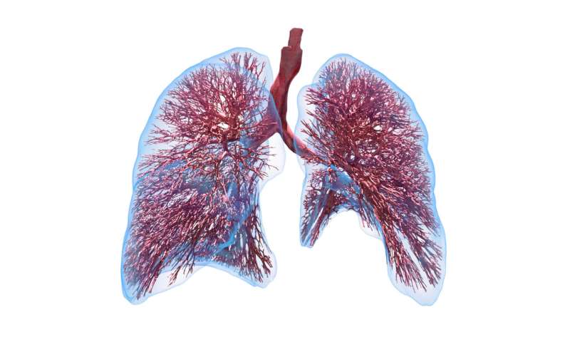
Although some medical texts still inaccurately portray the lung’s air sacs (alveoli) as similar to grapevines and bunches of grapes, lung tissue actually has a more sponge-like consistency. And it’s through this fine-walled tissue where the exchange between the air and the bloodstream occurs. Breathing comprises an extremely complex mechanical interaction between the different types of tissue, the liquid film on the tissue and the flow of air.
For several years, TUM researchers have been working to develop ever-more sophisticated models to simulate the behavior of lung tissue and airflow. Together with improved methods of micromechanical testing on lung tissue samples, their research has resulted in the creation of a computational lung model.
This model is the basis of a computer program that can calculate the local strains which would be placed on the lung’s microlevel tissues by different ventilator settings. Having these data at hand, medical staff and doctors can adjust the ventilator settings accordingly to provide a protective ventilation.
Using artificial intelligence to interpret the data
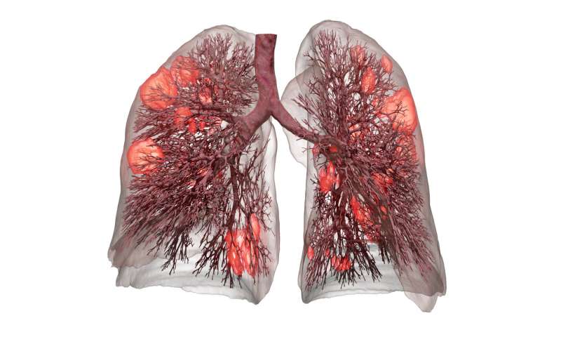
The current clinical standards guiding treatment with mechanical ventilation use a patient’s body weight to determine optimal ventilator pressure settings. However, the program developed by Prof. Wall and his team models the actual lung based on data compiled from a CT lung scan. It even considers the condition of individual areas of the lung that have already been damaged by the disease or previous injuries.
By measuring the changes in pressure and volume that occur during an inhalation and exhalation cycle, the digital lung model calculates the individual mechanical characteristics of the patient’s lungs. The result: a digital “twin” model of the patient’s lungs. It is so precise that it can accurately predict which ventilator settings will cause damage to the patient’s lungs.
From the research lab to the hospital—testing this model in real-life clinical settings
https://youtube.com/watch?v=KvzY5foV0oQ%3Fcolor%3Dwhite
Parallel to continuing his working group’s research together with clinical partners, Prof. Wall and three former colleagues founded the company Ebenbuild to bring their research into clinical practice as quickly as possible.
A key step in realizing this goal was automating the generation of lung models using artificial intelligence (AI). Prof. Wall and his team have harnessed the computing power of AI to developed a digital tool that can map a patient’s lungs and which can even be used for early detection of COVID-19 infections.
Source: Read Full Article