


Researchers in the United States have successfully delivered a critical structural protein found in severe acute respiratory syndrome coronavirus 2 (SARS-CoV-2) to a human cell line and tracked its localization deep within the cells.
The team says the development could help to further characterize key pathogenic mechanisms in SARS-CoV-2 infection and facilitate therapeutic approaches to coronavirus disease 2019 (COVID-19).
The researchers – from Arizona State University and Vanderbilt University in Nashville, Tennessee – delivered the SARS-CoV-2 envelope protein (S2-E) to human alveolar cells and tracked its location to key sites of coronavirus replication and assembly.
Charlies Sanders and colleagues say the ability to deliver S2-E to cells could lead to both chemical and biological approaches in future studies of SARS-CoV-2 pathogenesis and to the development of “Trojan Horse” antiviral therapies.
A pre-print version of the research paper is available on the bioRxiv* server, while the article undergoes peer review.
.jpg)
Understanding coronavirus pathogenesis is key to mitigating pandemics
Understanding the molecular underpinnings of coronavirus pathogenesis could mitigate both the current COVID-19 pandemic and potential future outbreaks.
Within SARS-CoV-2, the critically conserved S2-E protein has been shown to play an essential role in coronavirus assembly and budding.
A growing body of evidence suggests that S2-E is directly responsible for the acute respiratory distress syndrome (ARDS) that occurs in coronavirus infections. However, the protein’s role in viral pathogenesis is not well understood.
The S2-E protein is highly expressed in SARS-CoV-2 infected cells, but only a small fraction is incorporated into mature viral particles. This suggests the protein functions beyond its role as a mature structural protein, says Sanders and colleagues.
“Since SE-2 functions in multiple roles that are critical to viral fitness, it is desirable to develop methods to further characterize key pathogenic mechanisms,” writes the team.
What did the researchers do?
Sanders and colleagues set out to develop a robust method for delivering purified S2-E into cells, with the aim of enabling approaches to studying the protein’s function.
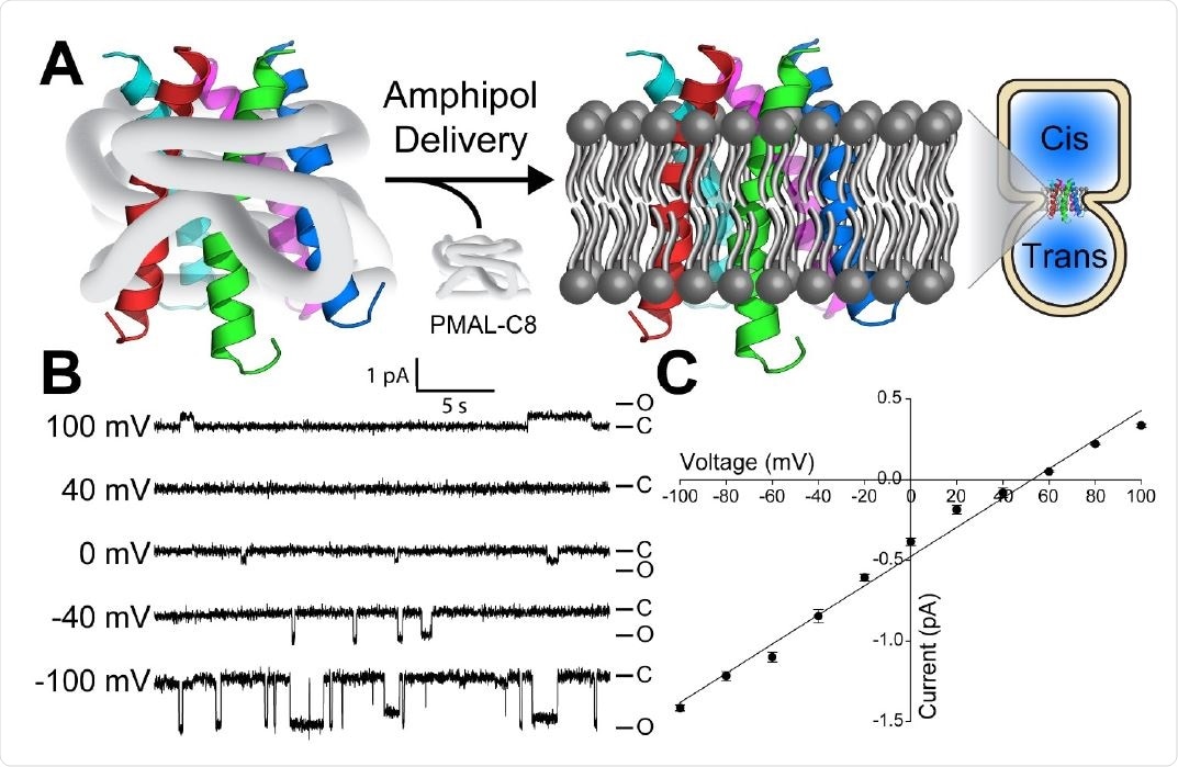
Now, the researchers have described the recombinant expression and purification of S2-E into amphipol solutions. Amphipols are a class of amphipathic polymers that can solubilize and stabilize native membrane protein folds, without disrupting the membranes.
First, the researchers showed that amphipol-based delivery of S2-E to pre-formed planar bilayers resulted in spontaneous membrane integration of the protein and the formation of ion channels.
Membrane capacitance measurements were performed to monitor the bilayer integrity during amphipol delivery.
This showed that not only was S2-E successfully inserted into the bilayers, but ion channel function was retained without significantly compromising bilayer integrity.
Delivering the protein to human alveolar cells
Next, the team demonstrated that amphipol-based delivery to SW1573 human alveolar cells (a COVID-19-relevant cell line) also resulted in membrane integration of S2-E.
The researchers then used monoclonal antibodies to pinpoint the final cellular location of S2-E. This revealed that the protein was concentrated in the area surrounding the Golgi and proximal to the cytosol-facing side of the endoplasmic reticulum-to-Golgi intermediate compartments (ERGIC).
Both the Golgi and the ERGIC are believed to be key sites of coronavirus replication and assembly, says Sanders and colleagues.
What are the implications of the study?
The researchers say the findings show that amphipol-based delivery can spontaneously insert S2-E into lipid bilayers to form ion channels. The protein can also be integrated into the plasma membrane of living human cells and subsequently trafficked to a location immediately adjacent to both the Golgi and ERGIC compartments.
The team says the approach could be used to deliver chemically modified full-length S2-E to cells in culture or possibly even to cells under physiological conditions.
“This capability enables a wide range of chemical and biological tools to explore the function of this protein or to test whether chemical-warhead-armed S2-E can play the role of a ‘Trojan horse’ to interfere with SARS-CoV-2 replication, potentially as an anti-COVID therapeutic or prophylactic,” says Sanders and colleagues.
“The results of this work also establish a general paradigm for using amphipols to deliver membrane proteins to living cells, although whether numerous other membrane proteins can be successfully delivered using this approach remains to be explored,” they conclude.
*Important Notice
bioRxiv publishes preliminary scientific reports that are not peer-reviewed and, therefore, should not be regarded as conclusive, guide clinical practice/health-related behavior, or treated as established information.
- Sanders C, et al. Delivery of recombinant SARS-CoV-2 envelope protein into human cells. bioRxiv, 2021. doi: https://doi.org/10.1101/2021.02.18.431684, https://www.biorxiv.org/content/10.1101/2021.02.18.431684v1
Posted in: Medical Research News | Disease/Infection News
Tags: Acute Respiratory Distress Syndrome, Antibodies, Cation, Cell, Cell Line, Coronavirus, Coronavirus Disease COVID-19, Horse, Ion, Ion Channel, Living Cells, Pandemic, Polymers, Protein, Research, Respiratory, SARS, SARS-CoV-2, Severe Acute Respiratory, Severe Acute Respiratory Syndrome, Structural Protein, Syndrome

Written by
Sally Robertson
Sally first developed an interest in medical communications when she took on the role of Journal Development Editor for BioMed Central (BMC), after having graduated with a degree in biomedical science from Greenwich University.
Source: Read Full Article