By Jayasheree Sundaram, MBA
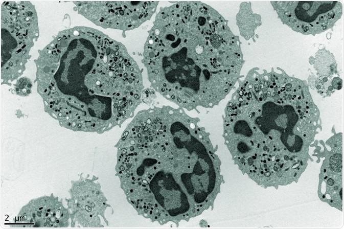
For single particle analysis, electron microscopy (EM) is critical. The resolution of EM allows the structure of a single molecule or compound to be imaged and elucidated.

Credit: Dlumen/Shutterstock.com
Noisy and disoriented two-dimensional (2D) images distort the structure of particles imaged using EM, but digital processing of these micrographs into clear three-dimensional (3D) images improves image quality. This is done computationally and follows a series of steps collectively termed as “single particle analysis techniques.”
Single particle analysis techniques
Two dimensional images are prepared through either negative staining or vitrification. These are then transformed into higher resolution, 2D projections that are then reconstructed to form a 3D structure.
The techniques involved in this transformation are:
2D image analysis
Processing the image
The initial image may need some corrections prior to 2D analysis. Common problems are given below.
- The images may be a blurred, as the sample has a tendency to move slightly during the process of imaging. They can also be blurred due to optics, where contrast transfer function (CTF) multiplies the ideal image and makes it complicated.
- There may be non-uniform brightness in the image, which happens because of either the thickness of the sample or uneven illumination
During the processing of images, instead of capturing images, a movie is recorded by the use of direct electron detectors, and then, the alignment of the movie frames is done computationally to average the frames together. To clear out irregular brightness of the images, high-pass filtering is carried out. Blurring due to optics is eliminated by estimating the CTF parameters and correcting them in the frequency domain.
Particle selection
The micrographs can be low contrast as well as can contain many contaminants or noise that should be ignored. Various semi-automated and automated methods are used for picking the particles out, such as template matching or selection of high-contrast regions.
Clustering
In this process, all images captured using EM are classified into different groups of similar images. Each group contains a set of images with similar view angles. The standard approach used in this method is k-means clustering, in which ‘k’ number of images are chosen as class exemplars and all other images are assigned to the most similar exemplar. A few other sophisticated methods are also available for clustering the images.
Class averaging
Averaging the images in each cluster helps in reducing the noise by canceling noises in different images. Based on the object that is sampled, there are two averaging techniques: electron crystallography (for periodic objects like 2D crystals) and single particle averaging (single molecules that are randomly oriented).
3D reconstruction
Image reconstruction
After clustering and averaging of images, the resultant images can be reconstructed to make a 3D structure. The primary step in this section can be performed two ways:
- When view angles are known: If the view angle of each of the particle is known, the easiest method to be followed is back-projection. In this process, each projection across the image is reversed back by a smear or coating. A blurred version of the real image is obtained through this, which can further be filtered with a high-pass filter. This is termed as filtered back-projection.
- When the view angles are unknown: If the view angles of the particles are not available, or unknown, yet there is a primary 3D model, then the process followed is 3D projection matching. Here, for each cluster or class average, the view angle that closely matches the 3D model is found out and by filtered back-projection of these estimated angles, the structure is reconstructed.
Framing
Used to frame or calculate an initial structure. Options include:
- Obtaining one from the previous experimental work
- Conducting lower-resolution specialized experiments
- Obtaining computational solutions directly through the following methods: common lines method and stochastic hill climbing
Acquiring atomic-resolution models
This is the final step involved in 3D reconstruction of micrographs. In this method, atomic-resolution models are fitted into EM structures of lower resolution.
Electron microscopy images that acquire atomic-resolution levels provide a powerful technique for the study of protein structures. These studies are carried out in low, medium, as well as high resolution. The resultant 3D data or information is most important and valuable for objects that are spherical. Examples of spherical objects include viruses or ribosome molecules.
Sources:
- https://www.ncbi.nlm.nih.gov/pmc/articles/PMC2777225/
- https://web.stanford.edu/class/cs279/lectures/lecture13.pdf
- http://www.jsbi.org/pdfs/journal1/GIW00/GIW00F15.pdf
- https://www.google.com/url?sa=t&rct=j&q=&esrc=s&source=web&cd=34&cad=rja&uact=8&ved=0ahUKEwiVmMT2gpLVAhUFjJQKHfaDBmQ4HhAWCC8wAw&url=http%3A%2F%2Fwww.biophys.mpg.de%2Ffileadmin%2Fuser_upload%2Flectures%2FEM%2FSingle_particle_image_processing.pdf&usg=AFQjCNHW1dG5o8xu92lvXwWZLym4uHl1hA
- http://events.embo.org/13-SAXS/presenter%27s_talks/Christiane%20Schaffitzel,.pdf
- http://www.astbury.leeds.ac.uk/ABSL/EM/CASE/single_virus.php
Further Reading
- All Single Molecule Experiments Content
- History of Single Molecule Experiments
- Single molecule detection of proteins in single cells
- Novel sensor selectively screens single molecules passing through nanopores
- Tunable Resistive Pulse Sensing
Last Updated: Aug 23, 2018
Source: Read Full Article


