Promising findings are emerging from a study coordinated by a research team of the University of Trento on medulloblastoma, the most common malignant brain tumor in children affecting the central nervous system. For the first time, scientists have grown organoids in the laboratory to simulate tumor tissue, and have identified the type of cell from which the tumor may originate.
The study was conducted by an international collaboration involving the research team led by Luca Tiberi of the Armenise-Harvard Laboratory of Brain Cancer at the Department of Cellular, Computational and Integrative Biology—Cibio of UniTrento, the Paris Brain Institute-Institut du Cerveau at Sorbonne Université in Paris, the Hopp Children´s Cancer Center (KiTZ) in Heidelberg, Germany, and Sapienza University in Rome. It was supported by Fondazione Armenise-Harvard, Fondazione Airc (Italian Association for Cancer Research) and Fondazione Caritro from Trento. The findings of the study, published in Science Advances, could lead to better and more effective treatments.
The team of researchers is proud of the results achieved. Luca Tiberi, coordinator of the study and corresponding author of the paper, comments, “For the first time, we have used an organoid model that we created and developed in past months. Thanks to these in vitro cancer tissues in 3D, we were able to identify the type of cell that can develop into medulloblastoma. These cells in fact express Notch1/S100b, and play a key role in the onset, progression and prognosis of this type of childhood brain cancer.”
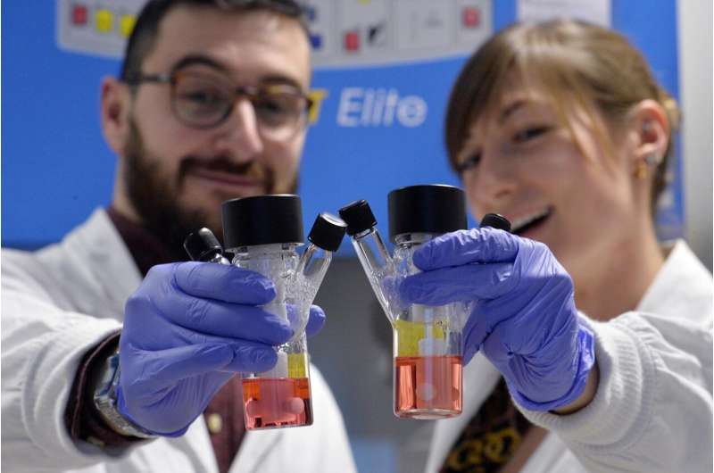
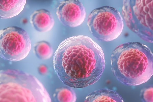
Organoids, grown by the hundreds in the laboratories of the University of Trento, are generated from skin or blood cells and look like irregular spheres the size of a peanut. Scientists use them to understand the genetic mechanisms responsible for the most common brain cancer affecting children and to explore new treatments for incurable conditions. Organoids were used to provide a model of the tumors in the laboratory. The findings may lead to advancements in brain cancer research, and in the future, could be used to study other tumors in a laboratory setting at a reduced cost, compared with previous technologies, and to conduct larger screenings to test new drugs and tailored treatments.
About organoids
Organoids are generated from skin or blood cells and look like irregular balls the size of a peanut. They are not cell aggregates, but specialized and organized cells that replicate as much as possible the organ under study.
These three-dimensional models allow scientists to do research in a laboratory setting. Working with patients in the case of the present study would be impossible, given that it focuses on brain cancer in children. That is why the team of researchers of the Armenise-Harvard Laboratory of the Department of Cellular, Computational and Integrated Biology—Cibio of the University of Trento used organoids to understand the genetic mechanisms of brain cancer in childhood and to find new treatments for these almost untreatable conditions.
Source: Read Full Article


