Nanomechanics is an emerging field that involves the study of mechanical properties, such as elastic, thermal, and kinetic properties of both biological and physical systems at the nanoscale.
Skip to:
- Mechanics in Biology
- Nanomechanical Properties of Biological Structures
- Globular Proteins
- Protein Filaments
- Isolated Cellular Structures
- Viruses
 molekuul_be | Shutterstock
Mechanics in Biology
The role of mechanical cues in biology is an emerging area of study, as it is being increasingly observed that mechanical cues play critical roles in many biological processes, such as cell differentiation, cell signaling, signal transduction, apoptosis, and in several disease states.
In several of these cases, mechanical cues may directly trigger the gene expression and signaling cascades that control the behavior of a cell or tissue. Thus, it is important to analyse the mechanical properties of participating proteins and protein networks. This can be done by studying proteins in stretched, bent, or loaded positions and understanding how their properties in terms of shape, size, and signaling may change in response.
Nanomechanical Properties of Biological Structures
Atomic force microscopy (AFM) is a tool commonly employed to explore the physical properties of structures in biology. In the method, a predetermined force is applied to the sample and the deformation that ensues due to this process is monitored and measured. This helps in understanding the three-dimensional molecular structure of the outer layer of a biological sample.
Globular Proteins
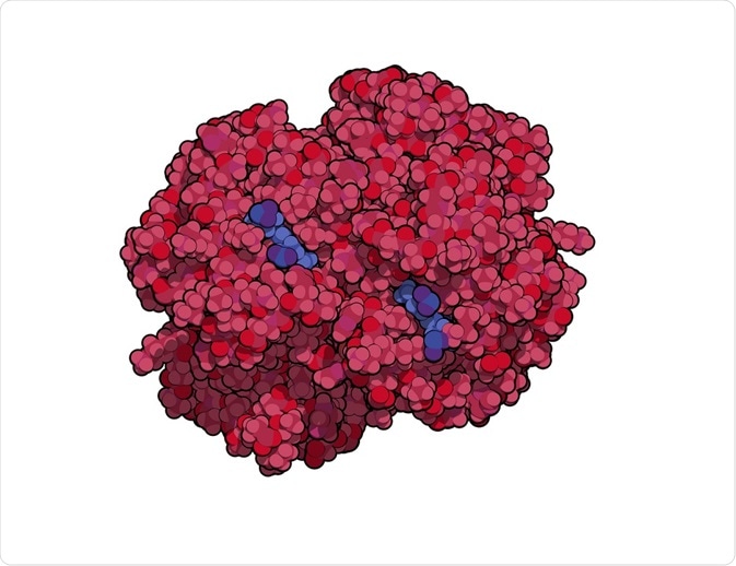
Atomic force microscopy has been used to study the mechanical properties of globular proteins. One of the first proteins analyzed using this method was lysozyme, which was the first enzyme structure determined using X-ray diffraction crystallography.
The method involved indenting the molecule and observing the deformation process to understand the underlying mechanical forces operating. Another such molecule was carbonic anhydrase, which consists of 259 amino acids that are folded into beta sheets.
This enzyme was compressed and stretched by atomic force microscopy and the data was validated using molecular modeling. However, in such cases, if the deformation is too high, it may lead to irreversible changes in the structure of the molecule.
Protein Filaments
Protein filaments are part of the cytoskeleton and also make up extracellular structures, including ligaments and tendons. Thus, understanding their mechanical properties can be very insightful. The cytoskeleton consists of actin, microtubules, and intermediate filaments.
An understanding of the mechanical properties of these filaments can generate insights on the global properties of cells. One of the first experiments to test the stiffness of microtubules was performed in 1995.
The shear modulus of microtubules was calculated to be 1.4 MPa. In its elastic state, microtubules are deemed to behave like a spring. Similarly, tau proteins are known to aggregate in some disease states such as Alzheimer's and other neurodegenerative disorders. Through an atomic force microscopy study, the binding of tau to microtubules was found to increase its elastic properties.
Isolated Cellular Structures
The height and volume measurements of chromosomes are determined using the three modes of atomic force microscopy: contact, tapping, and force mapping. In a study, the height and volume of human metaphase chromosome were measured. Also, the secretory products of a cell are stored inside secretory granules or vesicles.
Studies have investigated how mechanical properties determine the release of neurotransmitters from these vesicles. One of the methods that have been suggested is that the stiffness of the vesicles can indicate the process of exocytosis.
Viruses
Atomic force measurements of the mechanical properties of viral capsids have also been performed. These studies throw light on the infectivity and survival of the organism. In 2006, atomic force microscopy was used to determine the mechanical properties of wild-type and mutant strains of viruses.
Sources
- https://link.springer.com/content/pdf/10.1007%2Fs00424-008-0448-y.pdf
- http://web.mit.edu/cortiz/www/OrtizAbstractBioNanomechanics.pdf
- http://web.mit.edu/mbuehler/www/research/mechanobiology_May18.pdf
Further Reading
- All Nanoscience Content
- Characterization of Nanoparticles
- What are Nanomicelles?
- Applications of Nanomicelles
- Pioneering British Nanoscientists
Last Updated: May 24, 2019

Written by
Dr. Surat P
Dr. Surat graduated with a Ph.D. in Cell Biology and Mechanobiology from the Tata Institute of Fundamental Research (Mumbai, India) in 2016. Prior to her Ph.D., Surat studied for a Bachelor of Science (B.Sc.) degree in Zoology, during which she was the recipient of anIndian Academy of SciencesSummer Fellowship to study the proteins involved in AIDs. She produces feature articles on a wide range of topics, such as medical ethics, data manipulation, pseudoscience and superstition, education, and human evolution. She is passionate about science communication and writes articles covering all areas of the life sciences.
Source: Read Full Article


